MGF and PEG-MGF: the satellite-cell mechanism, and the absence of a human trial
MGF is the exercise-inducible splice variant of the IGF-I gene, and the C-terminal peptide it produces is a different molecule from systemic IGF-I. The mechanism is well described: it activates muscle satellite cells, its expression declines in ageing and fails in dystrophic muscle. The record cited here contains no human administration of PEG-MGF, and this page does not imply one.
29 October 2026 · 3 min read
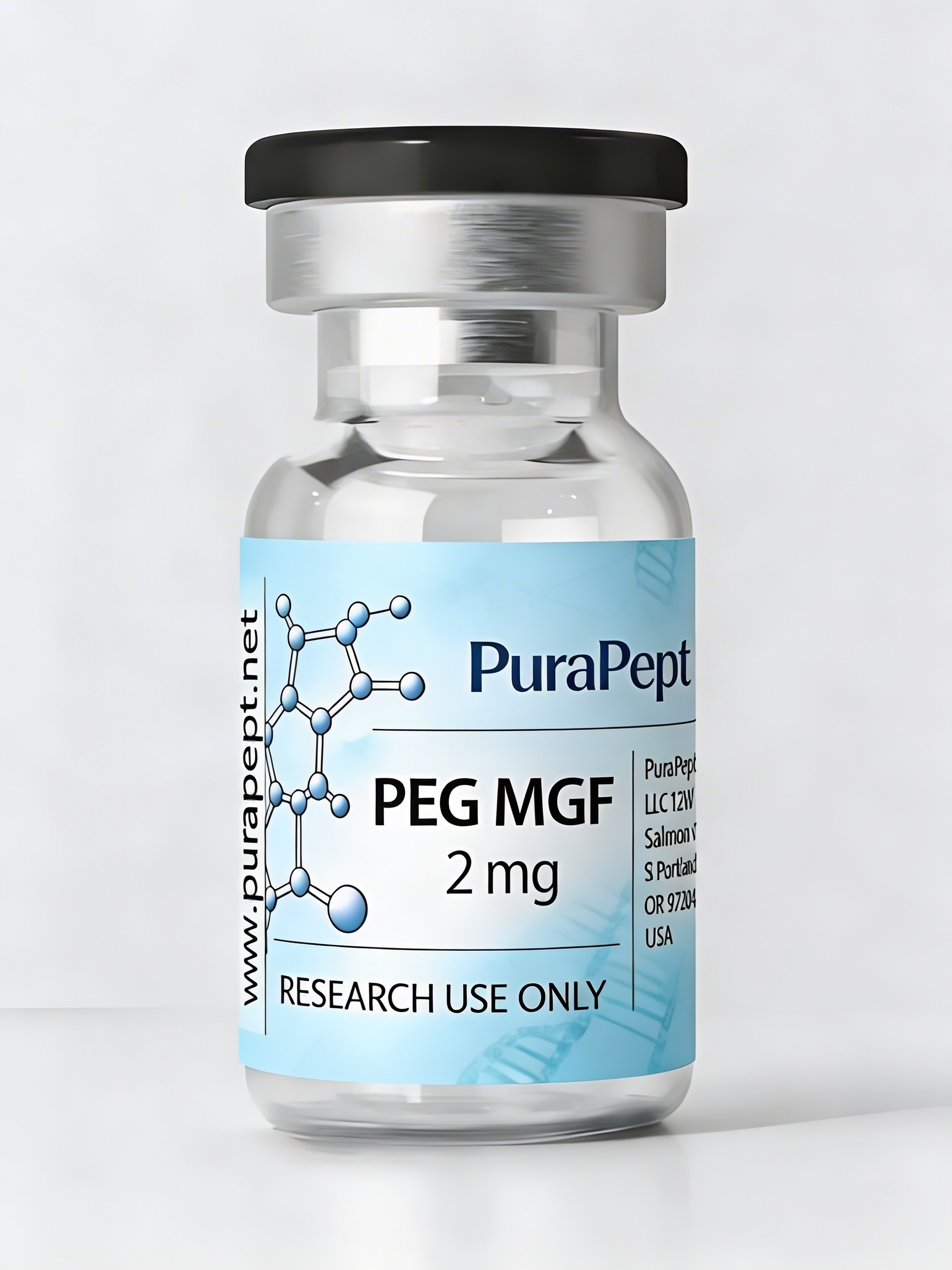 PEG MGF 2 mg
PEG-MGF is sold as an exercise mimetic. The mechanism that makes the claim plausible is real and well described, and this page reports it — then reports what is not there, which is the human administration record.
Why MGF is a different molecule from IGF-I
A 2003 review in the Journal of Muscle Research and Cell Motility explains the genetics. A group cloned three types of IGF-I in human muscle, all derived from the IGF-I gene by alternative splicing. One of them, an autocrine/paracrine splice variant, is only detectable after mechanical stimulation — that is MGF. The other, IGF-IEa, is systemic, produced by the liver and by other tissue including muscle.
The consequence is structural. As a result of a reading-frame shift, the MGF peptide has a different C-terminal sequence to IGF-IEa. The review states that the MGF C-terminal peptide “has been found to act as a separate growth factor and to initially activate mononucleated myoblasts (satellite cells).” MGF also responds to different signals and has different expression kinetics from IGF-IEa.
That last point is the one the exercise-mimetic framing usually omits. MGF is not IGF-I with a different label; it is a distinct C-terminal product of a reading-frame shift, with its own receptor behaviour and its own expression timing.
The satellite-cell activation, in a cell line
A 2010 Journal of Molecular Endocrinology paper gives a specific in-vitro result. Aged castrated rats were treated with testosterone or 5α-dihydrotestosterone, and during the first week DHT transiently increased soleus muscle nuclear density and induced expression of IGF1 and its splice variant MGF, without early regulation of MyoD, myogenin, monocyte nuclear factor or myostatin. A genome-wide microarray screen identified 70 candidate genes of 24,000 whose functions suggest initiation of remodelling and regeneration.
The MGF-specific part: treatment of C2C12 myoblasts with both IGF1Ea and the MGF C-terminal peptide increased nuclear beta-catenin in vitro. The proposed pathway is androgenic anabolism involving early downregulation of Axin and induction of IGF1, leading to nuclear accumulation of beta-catenin, a pro-myogenic, anti-adipogenic stem cell regulatory factor.
C2C12 is a mouse myoblast cell line. The half of the finding that concerns muscle repair in an intact animal is not in this experiment.
Why the ageing angle is the interesting one
The 2003 review states two limitations of the endogenous system that matter more than any performance claim. The mechanotransduction mechanism “may directly or indirectly involve the dystrophin complex, as dystrophic muscle, unlike normal muscle, is unable to express MGF in response to overload.” And the ability to express MGF has been found to decline markedly during ageing. The review’s conclusion is that the deficiency in expressing MGF and activating satellite cells in dystrophic and aged muscles may explain why muscle mass is not maintained in those situations, while in normal muscle MGF appears to initiate local muscle repair, with its overexpression resulting in hypertrophy.
So the case for MGF is strongest as an explanation of why older and dystrophic muscle repairs poorly — which is a physiological observation about the endogenous system, not a claim about administering the peptide.
What the record does not contain
Neither reference on this page administers PEG-MGF, MGF or any MGF fragment to a person, and neither reports a human outcome. There is no human pharmacokinetic profile, no human dose, no human route and no half-life for this compound in the records cited here. That is stated plainly rather than worked around, and it is the reason the page leads with mechanism and closes with limits.
What the data do not show
They do not show a human administration trial of PEG-MGF. The two records cited are a gene expression review and a rodent plus cell-line study.
They do not show muscle protein synthesis, lean mass or strength in people.
They do not show that satellite cells are activated in people by this compound, because the activation result printed here is in C2C12 mouse myoblasts.
They do not show that PEG conjugation extends a human half-life. No human half-life figure appears in either abstract.
One catalogue note, stated as a limit rather than as guidance: the preparation on this site is 2 mg. No abstract cited on this page prints a dose of any kind, human or animal, so this page cannot say what the shelf strength corresponds to in terms of exposure.
Where this sits on the shelf
The compound as supplied by the manufacturer: PEG MGF
What we supply
The PEG MGF preparation above, as supplied by the manufacturer, tracked, from inside the EU.
Research use only. This page summarises published studies for research reference. It is not medical advice, not a protocol, and not a suggestion for human use. Nothing we supply is for human or veterinary use.
References.
- Goldspink G. Gene expression in muscle in response to exercise. J Muscle Res Cell Motil. 2003;24(2-3):121-6.
- Gentile MA, Nantermet PV, Vogel RL, et al. Androgen-mediated improvement of body composition and muscle function involves a novel early transcriptional program including IGF1, mechano growth factor, and induction of {beta}-catenin. J Mol Endocrinol. 2010 Jan;44(1):55-73.